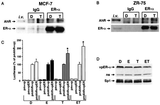
FIG. 8.
Activated AhR interacts with ERα. MCF-7 (A) and ZR-75 (B) cells were treated with DMSO (D) or TCDD (T) for 30 min. Nuclear extracts were isolated and immunoprecipitated with nonspecific mouse or goat IgG, anti-ERα (D12), or the AhR. AhR and ERα proteins were analyzed by Western blot analysis as described in Materials and Methods. In vitro-translated AhR and ERα were also used as markers. (C) Mammalian two-hybrid interactions of pm-AhR and vp-ER in ZR-75 breast cancer cells. ZR-75 cells were transfected with pGAL4, pm plus vp (empty vectors), pm plus vpER, pmAhR plus vp, or pmAhR plus vpER, treated with DMSO (D), 10 nM E (E), 10 nM (TCDD), or E plus TCDD (ET), and luciferase activity was determined as described in Materials and Methods. Results are expressed as means ± SE for three separate determinations for each treatment group. Luciferase activity significantly (P < 0.05) higher than that observed in cells transfected with pmAhR plus vp is indicated with an asterisk. (D) Levels of vpER were also determined by Western blot analysis in cells transfected with pmAhR and vpER. ns, nonspecific.

