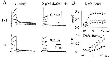
FIG. 2.
Impact of ERG1 B knockout on IK recorded for fetal mice. (A) Representative families of current traces of IK from fetal-day-18 ventricular myocytes from +/+ and ERG1 B−/− mice recorded before (left) and after (right) administration of 2 μM dofetilide. The Ic was elicited by depolarization to −30 to +50 mV in steps of 20 mV from a holding potential of −50 mV, and then their tail currents were recorded after returning to a potential of −50 mV. (B) The mean current-voltage relationships of dofetilide-sensitive (top) and -resistant (bottom) currents recorded from +/+ (black circle) and −/− (white circle) mice. Asterisks designate P values of <0.05.

