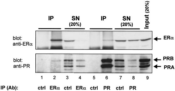
FIG. 1.
Interaction between ERα and PRB in T47D cells. Cell lysates were immunoprecipitated with anti-ERα or anti-PR antibodies or nonspecific IgG (control [ctrl]). SNs were collected, and IPs were removed from the beads in SDS sample buffer. IPs (lanes 1, 2, 5, and 6), SNs (lanes 3, 4, 7, and 8), and cell lysate (input) (lane 9) were analyzed by Western blotting with anti-ERα (upper panel) and anti-PR (lower panel) antibodies. ERα and PR bands are indicated by arrows. Lanes 3, 4, 7, and 8 correspond to 20% of the amount of proteins present in the supernatants. Lane 9 represents 20% of the amount of lysate used in the immunoprecipitation.

