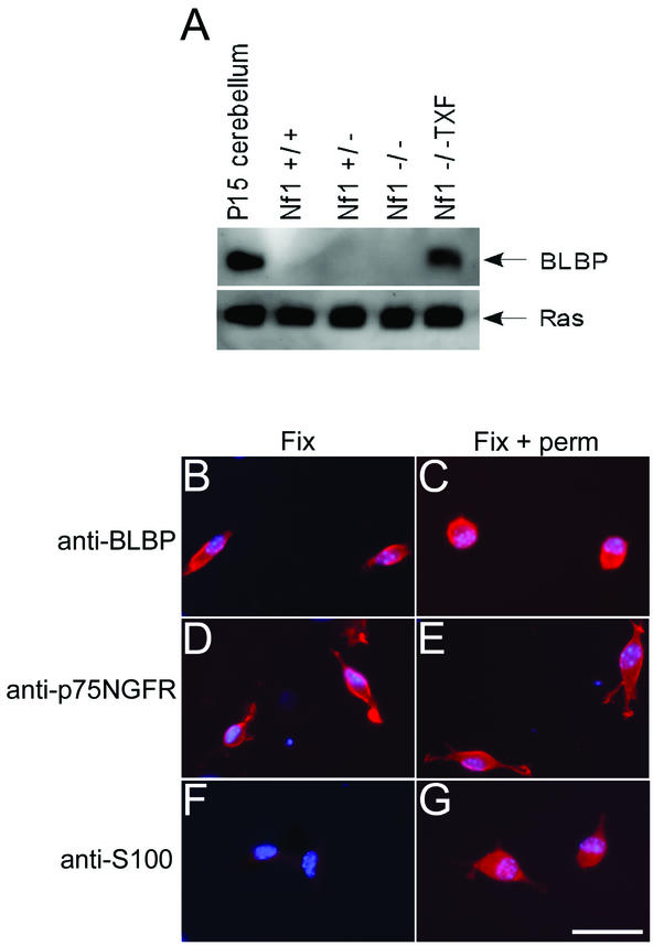
FIG. 2.
BLBP protein analysis in mouse Schwann cells. (A) Western analysis confirmed BLBP microarray data at the protein level. Cell lysates from wild-type, Nf1+/−, Nf1−/−, and Nf1−/− TXF Schwann cells were probed for BLBP expression. A 15-kDa protein corresponding to BLBP was detected in the Nf1−/− TXF cells only. Lysate from postnatal day 15 (P15) mouse cerebellum was used as a positive control. Blots were stripped and reprobed with anti-Ras antibody as a loading control. (B to G) Cells were fixed and immunolabeled with anti-BLBP, anti-p75NGFR, or anti-S100 antibodies, with (Fix + perm) or without (Fix) permeabilization of cell membranes. Labeling was detected with a rhodaminated secondary antibody (red). Cell nuclei were labeled with bis-benzimide (blue). BLBP is detected on the cell surface (B) and after permeabilization (C) in Nf1−/− TXF cells. The membrane protein p75NGFR is detected with (E) or without (D) permeabilization. The cytoplasmic S100 protein is detected only after permeabilization (F and G). The scale bar in panel G equals 50 μm and also applies to panels B to F.

