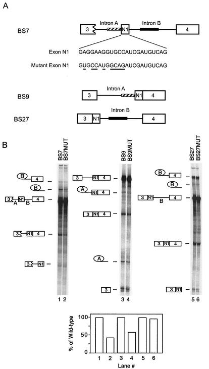
FIG. 1.
The N1 exonic enhancer element functions in vitro. (A) The pre-mRNAs used in the experiment are diagrammed. The nucleotides in exon N1 and mutant N1 exon are also shown, and the nucleotide changes in the mutant exon are underlined. The wild-type exon contains an inserted ClaI site 3′ of the mutation. This insertion has been previously shown not to affect N1 splicing (1a). The striped bar represents the upstream acceptor sequence, and the black bar represents the downstream enhancer sequence. (B) Splicing of pre-mRNAs with either a wild-type exon or a mutated exon in Weri-1 nuclear extract. Lanes 1 and 2, BS7 and BS7MUT; lanes 3 and 4, BS9 and BS9MUT; lanes 5 and 6, BS27 and BS27MUT. The products and intermediates are shown to the side of the gels. The percentage of RNA converted to mRNA was determined and compared between pairs of substrates, with the value for the mutant shown as a percentage of that for the wild-type.

