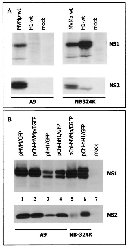
FIG. 4.
Expression of NS1 and NS2 proteins in A9 and NB-324K cells. (A) Cells were mock treated or infected at a MOI of 5 PFU/cell with wild-type MVMp (MVMp-wt) or H-1 (H1-wt) virus. At 18 h postinfection, cell cultures were metabolically labeled for 2 h with 200 μCi of Trans35S label (1,000 Ci/mmol; ICN Pharmaceuticals) in Met- and Cys-free Eagle's minimum essential medium supplemented with 5% dialyzed fetal calf serum. Cells were then lysed in modified radioimmunoprecipitation assay (MRIPA) buffer (10 mM Tris-Cl [pH 7.5], 150 mM NaCl, 1 mM EDTA, 1% Nonidet P-40, 0.5% sodium deoxycholate) containing a mixture of protease inhibitors (Complete; Roche Molecular Biochemicals). Equal amounts of labeled protein extracts (107 cpm) were immunoprecipitated with 10 μl of the polyclonal rabbit antiserum SP11 directed against the N terminus of NS1 and NS2 proteins of MVMp virus (3). The immunocomplexes were fractionated by electrophoresis on a sodium dodecyl sulfate-8% polyacrylamide gel and revealed by autoradiography. (B) A9 cells were transfected with 4 μg of plasmid DNA using the Lipofectamine transfection reagent as recommended by the supplier (Gibco BRL), while 25 μg of plasmid DNA was used to transfect NB-324K cells by the calcium phosphate precipitation method. Transfection efficiency was determined by the percentage of (E)GFP-positive cells and was identical for all the constructs. At 48 h after transfection, cells were lysed in MRIPA buffer, and 50-μg portions of protein extracts were analyzed by Western blotting. NS1 and NS2 were revealed by incubating the membrane with rabbit polyclonal antisera directed against the C-terminal part of MVMp NS1 (SP7) (13) and against the major isoform of MVMp NS2 (NS2p), respectively. The NS2p-specific antiserum was producedusing the keyhole limpet hemocyanin-coupled peptide AELGLRPEITWF. The membrane was further incubated with horseradish peroxidase-conjugated goat anti-rabbit antibody, and the immunoreactive proteins were visualized using Western Lightning Chemiluminescence Detection Reagent Plus (Perkin-Elmer Life Sciences).

