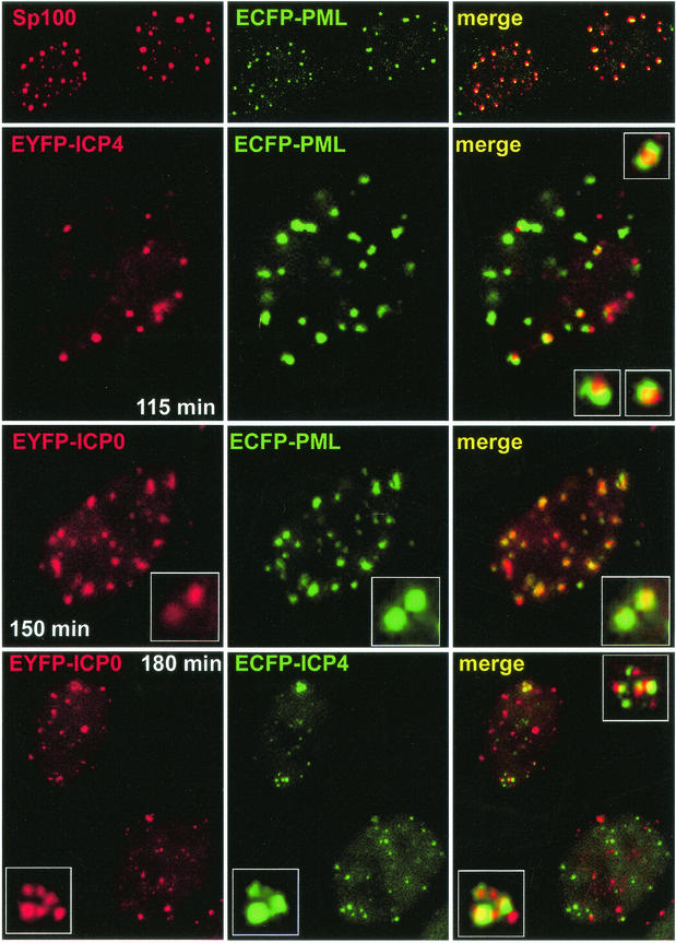
FIG. 4.
Examination of the relative locations of PML, ICP0, and ICP4 in live HEp-2 cells. Each row shows the single and merged channels of typical examples of cells from individual experiments. (Top row) ECFP-labeled PML (green) expressed from baculovirus Ac.CMV.ECFP-PML precisely colocalizes with endogenous Sp100 (red) in fixed cells as examined by confocal microscopy. (Row 2) HEp-2 cells infected the day before with Ac.CMV.ECFP-PML were infected with vEYFP-ICP4 (MOI = 10). The image was captured 115 min after addition of the virus (i.e., 55 min after a 60-min adsorption period). The inset images in the merged image show at higher magnification examples of ICP4 foci associated but not precisely colocalizing with PML. (Row 3) HEp-2 cells infected the day before with Ac.CMV.ECFP-PML were infected with vEYFP-ICP0 (MOI = 10). The image was captured 150 min after addition of the virus. Note that the colocalization of ICP0 and PML, indicated by the expanded insets, is far greater than that exhibited by ICP4 and PML. (Bottom row) HEp-2 cells were infected with virus vAFP4/0 (MOI = 10). The image was captured 180 min after addition of the virus. The inset images show examples of association but not precise colocalization of the ICP0 and ICP4 foci.

