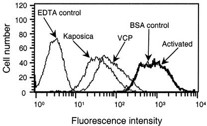
FIG. 3.
Inhibition of C3b deposition on erythrocytes during complement activation by kaposica and VCP. Classical pathway-mediated C3b deposition on erythrocytes was measured by incubating 5 μl of antibody-coated sheep erythrocytes (109/ml in GVB++: 5 mM barbital, 145 mM NaCl, 0.5 mM MgCl2, 0.15 mM CaCl2, and 0.1% gelatin, pH 7.4) with 1 μl of C8-deficient human serum (Calbiochem, San Diego, Calif.) and 44 μl of GVB++ or GVB++ containing 2 μM kaposica or VCP at 37°C for 30 min. Deposition of C3b was detected by fluorescence-activated cell sorting with fluorescein isothiocyanate-conjugated F(ab′)2 anti-C3 goat immunoglobulin G (Cappel Laboratories, Warrington, Pa.). Control samples contained either 10 mM EDTA or 2 μM bovine serum albumin (BSA).

