FIG. 6.
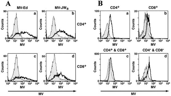
MV infects thymocytes from lck-SLAM tg mice. (A) In vitro MV infection of thymocytes from tg (open histogram) and non-tg (filled histogram) mice. Nonstimulated thymocytes were infected with MV-Ed (graphs a and c) or MV-JWB (graphs b and d) at an MOI of 1.0. CD4+ T cells (graphs a and b) and CD8+ T cells (graphs c and d) were stained for MV to detect viral proteins 2 days postinfection. (B) In vivo infection of hSLAM tg mice. Neonate animals of both non-tg (filled histogram) and tg (open histogram) mice were infected i.p. with MV-JWB at 4 × 10⁴ TCID50. Two days postinfection, CD4+ (graph a), CD8+ (graph b), CD4+ CD8+ (graph c), and CD4− CD8− (graph d) lymphocytes were analyzed for expression of MV proteins. Data shown are representative of three independent experiments.

