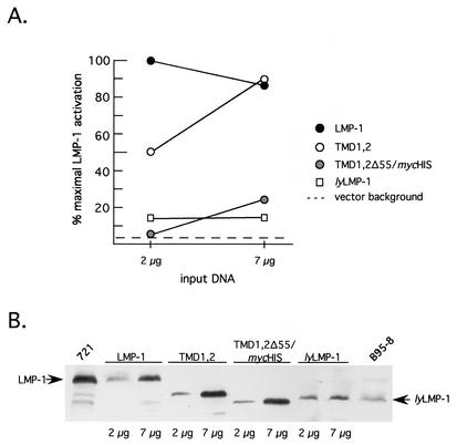
FIG. 4.
Activation of NF-κB by TMD1,2 in 293 cells. 293 cells were cotransfected with RSV-LMP-1 expression vectors encoding LMP-1, lyLMP-1, TMD1,2, or TMD1,2Δ55/mycHis (2 μg and 7 μg), together with an NF-κB-responsive luciferase reporter as described previously (4) and in Materials and Methods. (A) Extracts were assayed for luciferase activity 48 h posttransfection with a Dual Light assay kit (Tropix). Data are expressed as a percentage of maximal LMP-1 activation, with maximal activation by LMP-1 occurring at 2 μg of input DNA (423,310 relative light units [RLU]). The activation detected in the presence of 7 μg of empty vector control (pRC-RSV) is denoted by the dotted line. Solid circles, LMP-1; open circles, TMD1,2; shaded circles, TMD1,2Δ55/mycHis; open squares, lyLMP-1. (B) Extracts from A were assayed for LMP-1 expression by Western blot. Each lane was loaded with 5 × 10⁴ cell equivalents. The expressed protein is shown above the blot, and lanes are marked below the blot with input DNA amounts (2 μg or 7 μg). The amount of LMP-1 protein detected in each lane is representative of duplicate transfections in a single experiment. Arrows point to LMP-1 and lyLMP-1. 721 cells (5 × 10⁵ cells) and tetradecanoyl phorbol acetate- and butyrate-induced B95-8 cells (105 cells) (EBV-positive lymphoblastoid cell lines) were loaded on either side of the blot and served as markers for LMP-1 and lyLMP-1, respectively. Shown is a representative of three independent experiments. TMD1,2 activates NF-κB with a different concentration dependence than LMP-1 via a mechanism involving LMP-1 residues 331 to 386, which include CTAR2.

