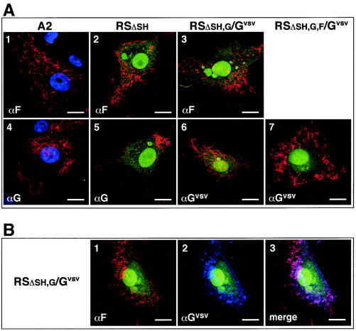
FIG. 9.
Cell surface localization of the Gvsv protein and HRSV G and F proteins in virus-infected cells by confocal microscopy. (A) Single antibody incubations for individual detection of G, F, and Gvsv. Vero cells were infected with the engineered viruses or virus A2 and fixed with freshly dissolved 4% paraformaldehyde at 24 h postinfection. G, F, and Gvsv proteins were detected by incubation with anti-VSIV G (αGvsv), anti-HRSV G (αG) or anti-HRSV F (αF) antibodies, followed by incubation with an Alexa-594 (red)-conjugated secondary antibody. A2-infected cells were, in addition, incubated with Hoechst stain to visualize nuclei (panels 1 and 4). Images were generated by sequential scanning for GFP expression (green) or Hoechst stain (blue) and expression of viral antigens (G, F, and Gvsv) (red). For each image, the antibody used is indicated in the lower left corner. Size bar, 20 μm. (B) Double antibody labeling for dual detection of F and Gvsv proteins. Vero cells infected with virus RSΔsh,g/Gvsv were fixed as described above and incubated with anti-HRSV F and anti-VSIV G antibodies simultaneously. The F protein was visualized with an Alexa-594 (red)-conjugated antibody, and Gvsv was visualized with an Alexa-350 (blue)-conjugated antibody. Images were generated by sequential scanning for GFP expression (green), F expression (red), and Gvsv expression (blue). The antibodies used are indicated. Panel 3 is a merged image of panels 1 and 2.

