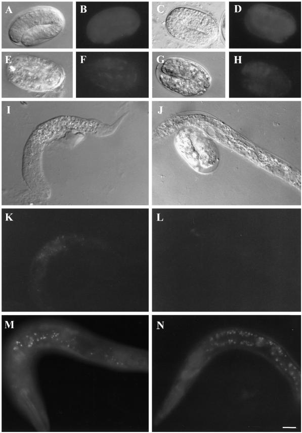
Figure 7.
RNAi of ς1 destabilizes both the UNC-101 and APM-1 proteins. The left panels show UNC-101::GFP expression and the right panels show APM-1::GFP expression. (A-H) show animals at their 2–3 fold embryonic stages, and (I-N) show animals at their L1 stage. A, C, E, G, I, and J are Nomarski images and all the others are GFP fluorescence images. (A-D) show the wild-type expression patterns of unc-101 and apm-1, and (E-L) show the results of RNAi of the ς1 gene. Both UNC-101::GFP and APM-1::GFP expression are reduced at the embryonic stage and the L1 stage by the RNAi as shown in F, H, K, and L. On the contrary, RNAi of ς2, the AP-2 small chain gene, neither reduced UNC-101::GFP nor APM-1::GFP expression as shown in M and N. Note that the nerve ring still contains APM-1::GFP expression even after RNAi of ς1 in L.

