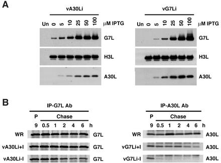
FIG. 10.
Stability of the A30L and G7L proteins. (A) BS-C-1 cells were either mock infected (lanes Un) or infected with vA30Li (left panel) or vG7Li (right panel) in the absence or presence of increasing concentrations of IPTG. After 24 h, cells were harvested and total cell lysates were analyzed by electrophoresis on an SDS-10 to 20% gradient polyacrylamide gel in Tricine buffer followed by Western blotting using antisera to the A30L, G7L, or H3L protein as indicated. (B) BS-C-1 cells were infected with VV WR, vA30Li, or vG7Li in the presence or absence of 50 μM IPTG. After 9 h, the cells were pulse-labeled with a mixture of [35S]methionine and [35S]cysteine for 15 min. Cells were either harvested immediately (lanes P) or incubated with excess unlabeled methionine for 0.5, 1, 2, 4, and 6 h (Chase lanes). Extracts of cells infected with vA30Li or vG7Li were immunoprecipitated with the G7L (IP-G7L Ab; left panel) or A30L (IP-A30C Ab; right panel) protein antiserum, respectively. The extracts from WR-infected cells were immunoprecipitated with G7L or A30L protein antiserum as indicated. The immunoprecipitated products were analyzed by SDS-PAGE and visualized by autoradiography.

