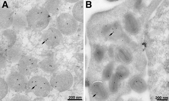
FIG. 4.
Localization of the G7L protein by immunoelectron microscopy. BS-C-1 cells were infected with VV WR at a multiplicity of infection of 2 PFU per cell. At 22 h, the cells were fixed in paraformaldehyde, cryosectioned, and incubated with an antibody to the peptide corresponding to amino acids 26 to 38 of the G7L protein and then with 10-nm-diameter gold particles conjugated to protein A. Electron micrographs are shown, with the scale indicated by the bars. (A) Field containing large numbers of IV; (B) field containing mature virions. Arrows point to representative gold particles.

