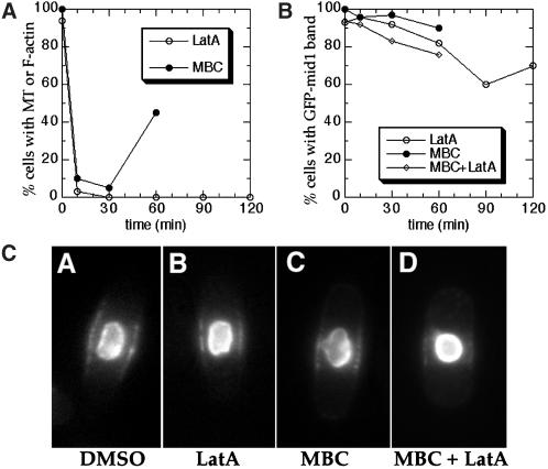
Figure 10.
Maintenance of mid1p broad band localization in the absence of F-actin and microtubule cytoskeletons. Wild-type cells carrying a nmt-GFP-mid1 construct (AP126 strain) were grown on minimal plates containing thiamine, inoculated into liquid cultures with thiamine, then treated with 200 μm LatA in 1% DMSO, 25 μg/ml MBC in 1%DMSO, both LatA and MBC in 2% DMSO, or 1% DMSO only. Samples were harvested at various time points after addition of drug. (A) Efficacy of the drug treatment. Open circles ○ represent percentage of LatA-treated cells with detectable F-actin structures as determined by rhodamine phalloidin. Filled circles ● represent percentage of MBC-treated cells with a microtubule (MT) over 2 μm in length. (n = 100 for each point). (B) Living cells with bright GFP-mid1 in the nucleus were assayed for a visible GFP-mid1 band by GFP fluorescence. All of the cells (100%) retained bright GFP-mid1 band staining after 1% DMSO treatment after 150 min. GFP-mid1-bands in cells treated with LatA were dimmer after 60 min. (C) GFP fluorescence images of representative AP126 cells 30 min after drug treatments.

