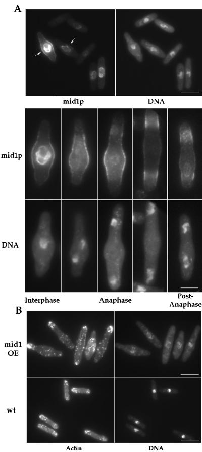
Figure 2.
Localization of mid1p and F-actin in mid1 overexpressing cells. (A) mid1p localization. Cells overexpressing mid1 grown at 30°C were fixed 22 h after thiamine removal from the medium and double stained with anti-mid1p antibody and DAPI. Interphase cells have nuclear and central cortical staining (arrows). In mitotic cells, nuclear staining disappears, and the cortical staining splits in two bands. Bars: top, 10 μm; bottom 5 μ m. (B) F-actin localization. Cells overexpressing mid1 induced for 22 h, and wild-type cells grown at 30°C were fixed and stained for F-actin with rhodamine phalloidin. Note increased concentration of actin patches at the cell center and the abnormal DNA staining in the mid1 overexpressing cells. Bars: 10 μm.

