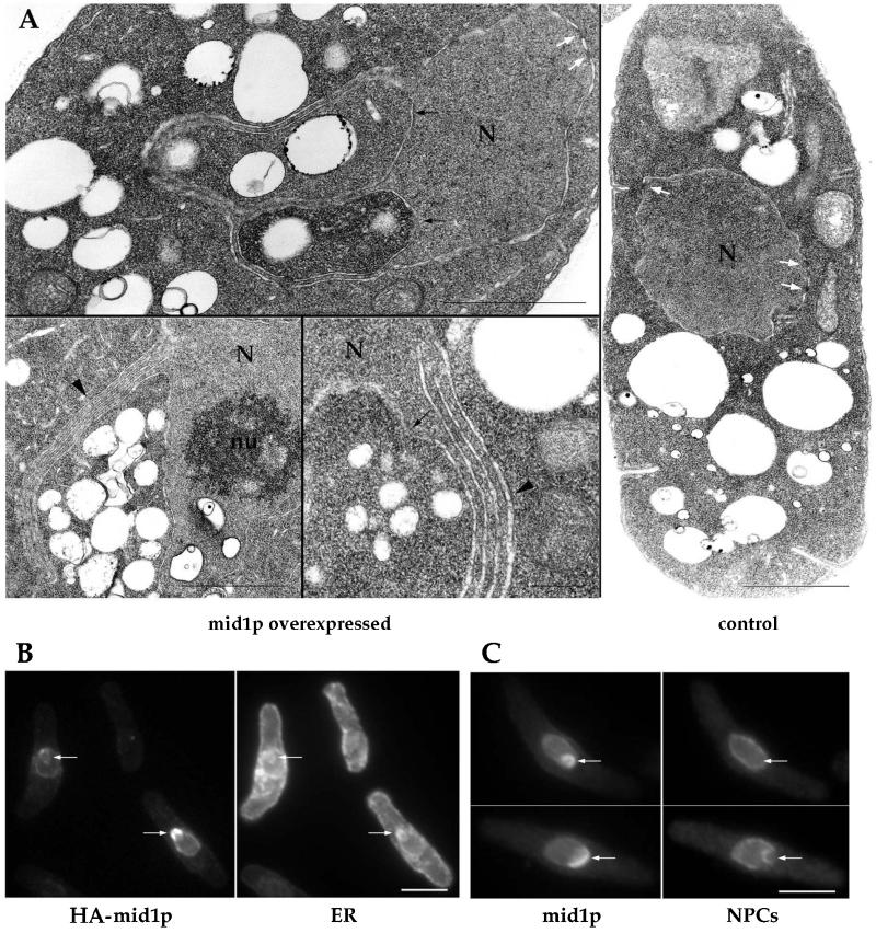
Figure 3.
Association of mid1p with NE/ER compartments and formation of karmellae-like structures. (A) Ultrastructural analysis of mid1 overexpressing cells. Cells overexpressing mid1 (24 h after thiamine removal) and control cells (grown in presence of thiamine) were fixed and processed for electron microscopy. Note invaginations of cytoplasm in the nucleus (black arrows) and karmellae-like multiple layers of nuclear envelope (black arrowheads). White arrows: nuclear pores. N: nucleus. nu: nucleolus. Bars: 1 μm, except bottom middle panel: 200 nm. (B-C) Colocalization of mid1p with the ER and nuclear pores. Cells overexpressing HA-tagged mid1p or mid1p were grown at 30°C, shifted to thiamine-free media for 22 h, and stained with mAb 3F10 (anti-HA) and anti-BiP antibody (ER marker) (B) or with the anti-mid1p antibody and mAb 414 (nuclear pore marker) (C). Note increased concentration of mid1p in regions that contain BiP but not nuclear pores (arrows). Bars: 5 μm.

