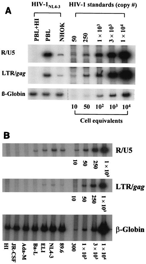
FIG. 1.
(A) Quantitative PCR analysis of HIV-infected NHOKs and PBLs. NHOKs were exposed to 400 ng of live HIV-1NL4-3 or heat-inactivated (HI) virus for 2 h at 37°C in the presence of 10 μg of Polybrene per ml. Cells were rinsed and cultured for 16 h prior to DNA extraction. Viral DNA was PCR amplified with the R/U5 primer pair (M667 and AA55, 140 bp) to detect early viral LTR reverse transcripts and LTR/gag (M667 and M661, 200 bp) for full-length reverse transcripts. The amplified products were resolved on a 6% nondenaturing polyacrylamide gel and visualized by autoradiography. Each lane represents DNA from approximately 5 × 10³ cells. β-Globin primers were used in parallel for a loading control. Heat inactivation of the virus was done for 1 h at 65°C, and activated PBLs were used as a positive control for virus infection. This is a representative result of more than five independent experiments. (B) Quantitative PCR analysis of NHOKs infected with primary HIV-1 strains.

