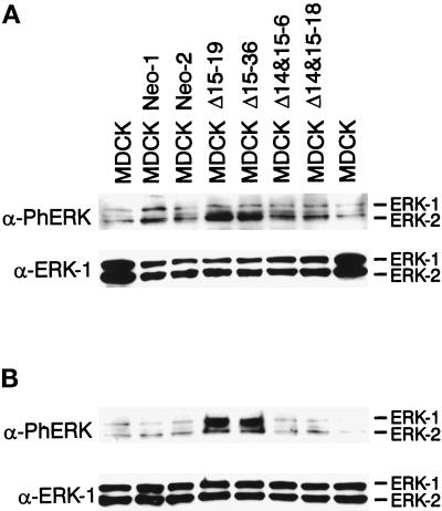
Figure 4.
Expression of the Δ15 PECAM-1 isoform in MDCK cells results in the activation of MAPK/ERKs. Cell extracts were prepared from parental or two representative clones of vector or PECAM-1 transfected cells grown under normal conditions (A) or serum starved for 48 h followed by 10 min of serum stimulation (B). Equal amounts of protein (30 μg) were analyzed by SDS-PAGE and Western blotting with either antiphospho-MAPK/ERKs (upper panels) or anti-ERK-1 (lower panels). Note the increased levels of constitutive (A) and serum-stimulated (B) phosphorylated active MAPK/ERKs in the Δ15 PECAM-1–expressing cells. These experiments were repeated three times with identical results.

