Figure 6.
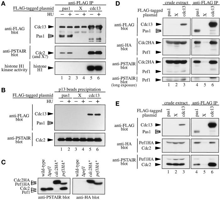
Pas1 cyclin associates in vivo with both Cdc2 and Pef1 kinases. (A) A Cdc2 kinase-like protein coprecipitates with Pas1 cyclin. Lysates were prepared from Δpas1 cells (K182-A7) expressing pREP1-FLAGpas1 (lanes 1 and 2), pREP1-X (lanes 3 and 4), or pREP1-FLAGcdc13 (lanes 5 and 6). Cells were cultured in MM (+N/2%G) in the absence (lanes 1, 3, and 5) or presence (lanes 2, 4, and 6) of 12 mM hydroxyl urea. Lysates were subjected to immunoprecipitation with the anti-FLAG M2 antibody. Immunoprecipitates were immunoblotted with the anti-FLAG M2 (top panel) or anti-PSTAIR (middle panel) antibodies, respectively, or assayed for histone H1 kinase activity (bottom panel). FLAG-Cdc13 protein comigrated with IgG (top panel; lanes 5 and 6). (B) The Pas1 cyclin-associated kinase does not bind Suc1p. The same lysates used in A were incubated with p13suc1 beads to pull down a Cdc2 kinase complex. Precipitates were immunoblotted with anti-FLAG M2 (upper panel) or anti-PSTAIR (lower panel) antibody. (C) Identification of Cdc2p and Pef1p molecules. Lysates were prepared from wild-type (EV3A), Δpef1 (K571-2D), cdc2HA+ (K230-A6), and pef1HA+ (K566-11) cells. The whole cell lysates were separated by SDS-PAGE and immunoblotted with anti-PSTAIR (left panel) or anti-HA (right panel) antibodies. (D and E) Pas1 cyclin associates in vivo with Cdc2 and Pef1 kinases. (D) Lysates were prepared from cdc2HA+ cells (K230-A6) expressing pREP1-FLAGpas1 (lanes 1 and 4), pREP1-X (lanes 2 and 5), or pREP1-FLAGcdc13 (lanes 3 and 6). The lysates were immunoprecipitated with the anti-FLAG M2 antibody. The whole cell lysate (left panels; lanes 1–3) and immunoprecipitates (right panels; lanes 4–6) were immunoblotted with the anti-FLAG D-8 (top panels), anti-HA (second panels), or anti-PSTAIR (third and bottom panels) antibody. In this experiment, an anti-FLAG D-8 rabbit polyclonal antibody was used to avoid an undesired reaction with mouse IgG (top panels). (E) Lysates were prepared from pef1HA+ cells (K566-11) expressing pREP1-FLAGpas1 (lanes 1 and 4), pREP1-X (lanes 2 and 5), or pREP1-FLAGcdc13 (lanes 3 and 6). The lysates were immunoprecipitated with the anti-FLAG M2 antibody. The whole cell lysate (left panels; lanes 1–3) and immunoprecipitates (right panels; lanes 4–6) were immunoblotted with the anti-FLAG D-8 (top panels), anti-HA (middle panels), or anti-PSTAIR (bottom panels) antibody. The FLAG-tagged proteins were detected by the anti-FLAG D-8 rabbit polyclonal antibody to avoid undesired reaction with mouse IgG (top panel).

