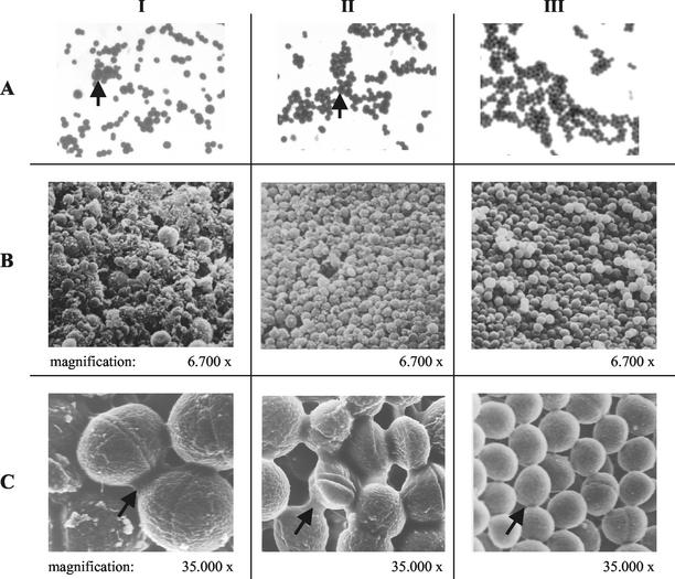
FIG. 2.
Gram staining and SEM of fried-egg SCVs (panels I), pinpoint SCVs (panels II), and normal S. aureus (panels III). (A) Gram staining. Arrows indicate large cocci. (B and C) Low- and high-magnification SEM, respectively. Arrows indicate the enhanced intercellular substance present in SCVs compared to normal S. aureus.

