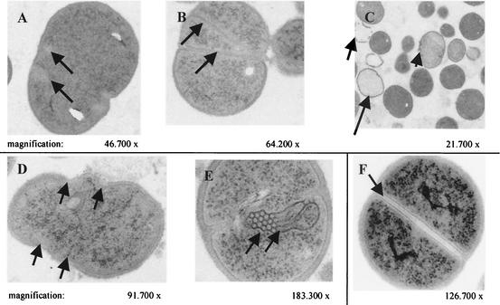
FIG. 3.
TEM of fried-egg SCVs (A, B, and C), pinpoint SCVs (D and E), and normal S. aureus (F). (A and B) Arrows indicate incomplete or multiple cross walls. (C) Incomplete cross wall (small arrows), cellular debris (intermediate arrow), and empty cells (large arrows) are seen. (D and E) Pinpoint SCVs with atypical unrounded cells and mesosome-like structures (arrow). (F) Normal S. aureus cells displaying regular cell separation by a cross wall (arrow) surrounding a highly contrasting splitting system.

