Figure 1.
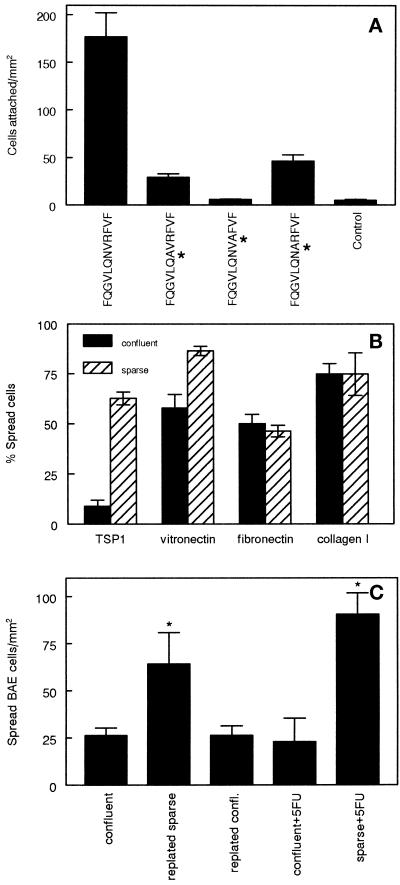
Spreading of BAE cells on TSP1 is regulated by confluence but not by proliferation. (A) Adhesion of endothelial cells on an α3β1 integrin–binding peptide from TSP1. TSP1 peptide 678 (FQGVLQNVRFVF) or analogues of this peptide with the indicated Ala substitutions (*) were adsorbed on bacteriological polystyrene substrates at 10 μM in PBS. Direct adhesion of BAE cells to the adsorbed peptides or uncoated substrate (control) is presented as means ± SD (n = 3). (B) Loss of cell–cell contact stimulates endothelial cell spreading on TSP1. Two flasks of BAE cells were grown to confluence. One flask was harvested and replated in fresh medium at 25% confluence. Fresh medium was added at the same time to the second flask. After 16 h, cells from both flasks were dissociated with the use of EDTA, and adhesion was measured on substrates coated with 40 μg/ml TSP1, 10 μg/ml vitronectin, 20 μg/ml plasma fibronectin, or 5 μg/ml type I collagen. The percent spread of cells from confluent (closed bars) or sparse cultures (striped bars) after 60 min is presented as means ± SD (n = 3) for a representative experiment. (C) Cell contact–dependent regulation of α3β1 integrin is independent of proliferation. Confluent BAE cell cultures and duplicate cultures treated with 5-fluorouracil were fed (confluent and confluent+5FU), dissociated and replated at low density in fresh medium 24 h before use (replated sparse and sparse+5FU), or dissociated and replated at their original density in fresh medium for 24 h (replated confl.). After 24 h, the cells were dissociated and tested for spreading on TSP1. Results are presented as means ± SD (n = 3). Significance was assessed with the use of a two-tailed t test, and values of p < 0.05 compared with the confluent control are indicated by asterisks above the bars.

