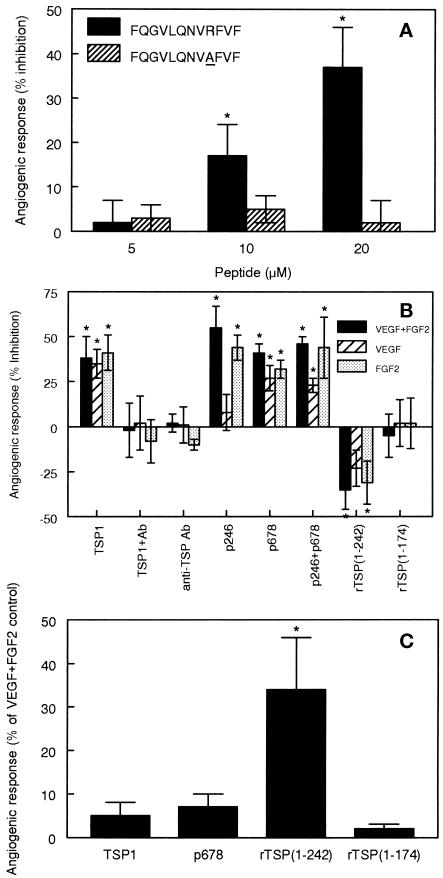
Figure 11.
Modulation of chick CAM angiogenesis by the α3β1-binding sequence in TSP1. (A) The TSP1 α3β1 integrin–binding peptide inhibits angiogenesis. Polymerized collagen gels containing the angiogenic growth factors VEGF and FGF2 in the presence or absence of the indicated concentrations of the TSP1 peptide FQGVLQNVRFVF (peptide 678; closed bars) or the control peptide FQGVLQNVAFVF (peptide 690; striped bars) were placed on the outer one-third of 10-d chick CAMs for 24 h. Each CAM contained two pellets for each peptide concentration as well as positive and negative controls. The ability of the peptides to modulate growth factor–driven angiogenesis was assessed by injection of FITC-dextran and digital image analysis. The percent inhibition relative to controls is presented as means ± SD for each group (n = 8). Conditions that significantly differed from their respective controls based on a two-tailed t test with p < 0.05 are marked with asterisks. (B) Inhibition of CAM angiogenesis stimulated by VEGF and FGF2(closed bars) or by VEGF (striped bars) or FGF2 alone (shaded bars) was assessed as in A in the presence of the indicated effectors: 10 μg of TSP1, 10 μg of TSP1 plus 25 μg of anti-TSP1, 25 μg of anti-TSP1 (Ab), 20 μM peptide 246 (KRFKQDGGWSHWSPWSS from the type 1 repeats of TSP1), 20 μM peptide 678, 20 μM peptide 246 plus peptide 678, or 20 μg of recombinant TSP1 fragments containing residues 1–242 or 1–174 of the mature protein. Results are presented as means ± SD (n = 3–9). (C) Direct stimulation of angiogenesis by recombinant TSP1 fragments. Angiogenesis in the absence of growth factors was determined in the presence of 10 μg of TSP1, 20 μM peptide 678, or 20 μg of recombinant TSP1 (residues 1–242 or 1–174). Results are presented as percent of the positive control stimulated by VEGF plus FGF2 (means ± SD, n = 4–9).

