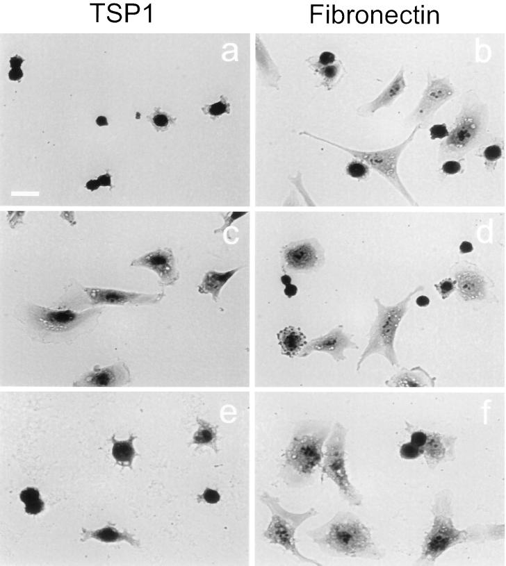
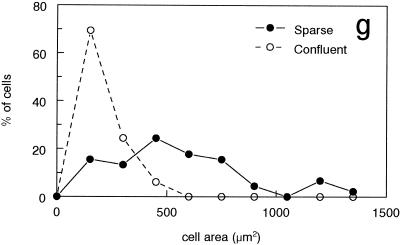
Figure 2.
Spreading on TSP1 induced by loss of cell–cell contact is inhibited by the α3β1 integrin–binding peptide from TSP1. BAE cells dissociated with the use of EDTA from confluent (a and b) or 16-h sparse cultures (c–f) were incubated for 60 min on substrates coated with 40 μg/ml TSP1 (a, c, and e) or 20 μg/ml fibronectin (b, d, and f). Adhesion was performed in the presence of 30 μM TSP1 peptide 678 (e and f). Cells were fixed with 1% glutaraldehyde and stained with the use of Diff-quik (Dade, Miami, FL). Bar in a, 25 μm. (g) Spread of cell areas for 100 cells each from the experiments presented in a (○) and c (●) were quantified from digitized images with the use of Image-Pro Plus software.


