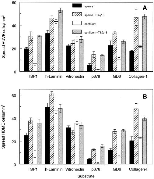
Figure 3.
Cell contact specifically regulates spreading of human endothelial cells on α3β1 integrin ligands. (A) HUVE cells harvested from sparse (closed and striped bars) or confluent cultures as in Figure 1 (open and gray bars) were plated on substrates coated with 10 μg/ml TSP1, 10 μg/ml human placental laminin, 5 μg/ml vitronectin, 5 μM TSP1 peptide 678, 5 μM laminin-1 peptide GD6 (KQNCLSSRASFRGCVRNLRLSR), or 5 μg/ml type I collagen. The cells were suspended in medium 199/0.1% BSA (closed and open bars) or the same medium containing 5 μg/ml β1 integrin–activating antibody TS2/16 (striped and gray bars). The number of spread cells was determined at 60 min and is presented as means ± SD (n = 3). (B) HDME cells were treated as in A.

