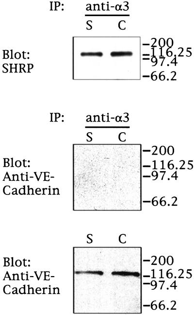
Figure 6.
α3β1 integrin and VE-cadherin expression in endothelial cell cultures. HUVE cells grown under sparse (S) or confluent (C) conditions were biotinylated, immunoprecipitated with the use of anti-α3 integrin antibody (P1B5), and fractionated on 10% SDS-polyacrylamide gels (upper and middle panels) along with equal amounts of proteins from total cell lysates (10 μg; lower panel). Surface proteins were detected with the use of HRP–streptavidin (SHRP) and enhanced chemiluminescence. VE-cadherin in the α3 immunoprecipitate (middle panel) or total lysate (lower panel) was detected by blotting. Migration of molecular weight markers is indicated on the right.

