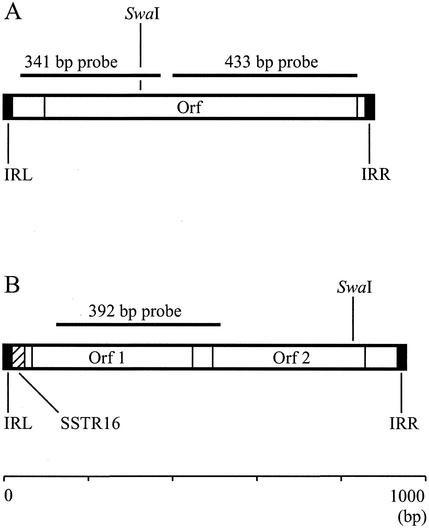
FIG. 1.
Organizations of ISs ISFtu2 (A) and ISFtu1 (B). Terminal inverted repeat sequences to the left (IRL) and right (IRR) are depicted as closed bars; an internal SSTR16 in ISFtu1 is depicted as a hatched bar. The locations of open reading frames (Orfs), the DNA probes used, and the recognition site for the restriction enzyme SwaI are indicated.

