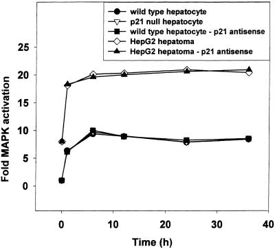
Figure 1.
Treatment of hepatocytes and hepatoma cells expressing ΔB-Raf:ER with 4-hydroxytamoxifen causes activation of MAPK. Cells were infected with kinase active ΔB-Raf:ER poly-L-lysine adenovirus (at 250 multiplicity of infection (m.o.i.), followed by culture as in Methods. As indicated, and in addition to ΔB-Raf:ER infection, cells were also infected with either null recombinant adenovirus or a recombinant adenovirus expressing p21 antisense mRNA, (at 100 m.o.i. each). After 24 h to allow protein/mRNA expression, hepatocytes were treated with either vehicle control or with 100 nM 4-hydroxytamoxifen for 36 h (total time in culture 60 h). Cells were assayed for MAPK activity before 4-hydroxytamoxifen addition (0 min), 1 h, 6 h, 12 h, 24 h, and 36 h after addition of 4-hydroxytamoxifen. Data are the means of triplicate determinations from 3 separate experiments/animals (to the nearest 50 cpm ± SEM) and are expressed as cpm incorporated into myelin basic protein above background (250 ± 50 cpm) in the standard MAPK assay (Methods). No activation of MAPK was observed in cells infected with kinase inactive ΔRaf:ER (301) treated with 4-hydroxytamoxifen (our unpublished observations). No activation of MAPK was observed in cells infected with ΔB-Raf: ER treated with matched vehicle control (DMSO) (our unpublished observations). Addition of 50 μM PD98059 to the culture media abolished the activation of MAPK by ΔB-Raf:ER in all of the cells examined above (our unpublished observations), in agreement with Auer et al. (1998b).

