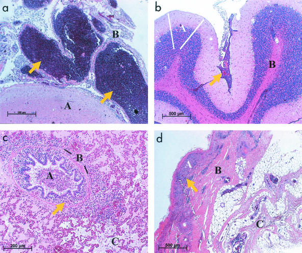
FIG. 1.
Histology of porcine malignant catarrhal fever (hematoxylin-eosin). (a) Cerebrum, pig 1, showing severe nonpurulent meningoencephalitis. Arrows point to massive accumulations of round cells in the meninges. A, grey matter (cortex cerebri); B, meninges (pia mater). Bar, 500 μm. (b) Cerebellum, pig 2. Arrow points to disseminated round cell infiltration in the meninges, indicating severe nonpurulent meningoencephalitis, although not as severe as in panel a. A, grey matter (cortex cerebri), spanned by white lines; B, white matter (corpus medullare); C, meninges (pia mater). Bar, 500 μm. (c) Lung, pig 5. Catarrhalic bronchopneumonia with purulent bronchitis, bronchiolitis, edema, peribronchitis, and peribronchiolitis with round cell cuffs. Arrow points to an accumulation of round cells in the area of a bronchiolus. A, lumen of bronchiolus; B, plain muscle cells (lamina muscularis mucosae), emphasized by black lines; C, destroyed alveoli. Bar, 200 μm. (d) Skin, pig 3. Alterations include hyperkeratosis, ulceration, and perivascular dermatitis with lymphocytes and plasma cells. Arrow points to accumulation of round cells. A, epidermis; B, cutis; C, subcutis. Bar, 500 μm.

