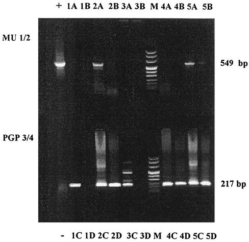
FIG. 1.
Typical nested PCR amplification findings. Following the first (top) and second (bottom) rounds of PCR amplification with primer pairs MU1-MU2 and PGP3-PGP4, respectively, 15 μl of sample was applied to a 1.6% agarose gel for electrophoretic separation. +, positive control; −, negative control after MU1-MU2 and PGP3-PGP4. Lanes 1A and 1C, spiked samples of sample 1; 1B, sample 1 after MU1-MU2 stage; 1D, sample 1 after PGP3-PGP4 stage. Samples 2 to 5 are categorized in the same way. Lane M, DNA molecular size markers. After PCR, sample 1 was negative, sample 2 was positive, 3 was negative, and samples 4 and 5 were positive.

