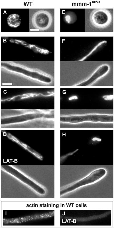
Figure 3.
Mitochondrial morphology is altered in mmm-1RIP mutants. Neurospora cells of wild-type (A–D) and mmm-1RIP23 (E–H) were stained with the mitochondria-specific vital dye DiOC6 and observed by fluorescence and phase contrast microscopy. (A, E) Conidia. (B, D, F, H) Hyphal tips 1 h after germination. (C, G) Hyphal cells 6 h after germination. (D) Hyphal tip of wild-type treated with 20 μg/ml LAT-B for 30 min. (I, J) Wild-type cells that were mock treated (I) or treated with 20 μg/ml LAT-B for 30 min (J) were subjected to indirect immunofluorescence to localize actin. Note that the image of the LAT-B–treated sample was taken with a 5 times longer exposure than that of the mock-treated cells. Bars, 5 μm.

