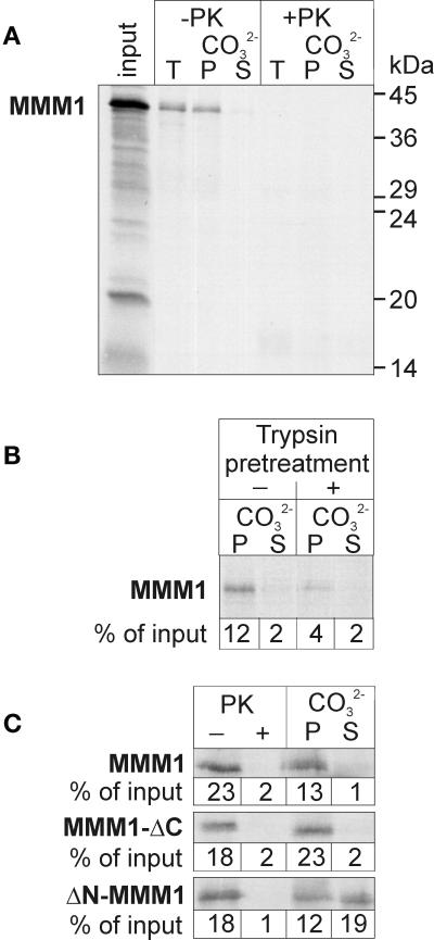
Figure 5.
In vitro import of MMM1. (A) Insertion of imported MMM1 into the outer membrane. MMM1 was synthesized in the presence of [35S]methionine and incubated with isolated mitochondria for 20 min at 20°C. After import, organelles were subjected to flotation in a sucrose gradient. Mitochondria were then either treated with 100 μg/ml proteinase K (+PK) for 15 min on ice or left untreated (−PK). Half of the reisolated organelles was directly precipitated with trichloroacetic acid (total, T), whereas the other half was resuspended in 0.1 M Na2CO3 and separated into pellet (P) and supernatant (S) fractions. The input lane shows 40% of the radiolabeled material added to the import reactions. Molecular size markers are indicated at the right. Proteins were analyzed by SDS-PAGE, blotting to nitrocellulose, and autoradiography. (B) Receptor dependence of MMM1 import. Isolated mitochondria were either pretreated with 40 μg/ml trypsin for 15 min on ice to cleave import receptors (+trypsin pretreatment) or left untreated (−trypsin pretreatment). Then, import, flotation of mitochondria and carbonate extraction were performed as in A. Import of MMM1 was quantified by densitometry and is indicated as percentage of input, where 100% represents the total radioactivity added to each import reaction. (C) Import of truncated MMM1 proteins. MMM1 and two truncated versions, MMM1-ΔC and ΔN-MMM1, were imported into mitochondria as in A. After flotation, mitochondria were treated with 100 μg/ml proteinase K (+PK) for 15 min on ice or left untreated (−PK), or were resuspended in 0.1 M Na2CO3 and separated into P and S fractions. Import was quantified by densitometry and is indicated as percentage of input.

