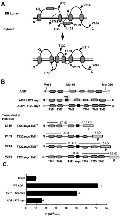
Figure 1.
(A) Diagram of initial AQP1 four-spanning ER topology and mature six-spanning plasma membrane topology. Shaded ovals represent TM segments and open circles represent the glycosylation site at residue N42. Curved arrows indicate the predicted 180-degree rotation of TM3 required for maturation from the four-spanning to the six-spanning structure. Straight arrows indicate the site and topological orientation of the P reporter for fusion proteins at indicated truncation sites. (B) Scheme of epitope-tagged AQP1 constructs indicating the relative locations of myc and P reporters. Black rectangles represent the myc epitope tag and hatched rectangles represent the P reporter. Predicted masses of myc-tagged truncated polypeptides are indicated. (C) Water permeability (Pf) of oocytes injected with water or cRNA encoding wild-type AQP1, AQP1.T120.myc, and AQP1.P77.myc. Results represent the average of two separate experiments, six oocytes per group, mean ± SE.

