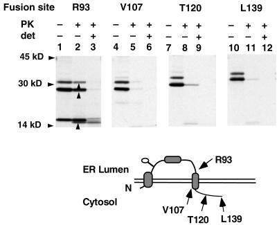
Figure 2.
Cotranslational topology of TM3 in XO. AQP1 was truncated and fused to the C-terminal P reporter at residues R93, V107, T120, and L139 as indicated. Oocytes expressing in vitro transcribed cRNA were homogenized, digested with PK, and immunoprecipitated with anti-prolactin antiserum. Upward arrowheads (lane 2) indicate PK-protected, P-reactive fragments. The topological location of the P reporter in each fusion protein is shown in the diagram beneath the autoradiograms. In lane 3, a small amount of protease-resistant material was recovered in the presence of detergent. This is observed in some experiments and likely represents protein that, for unclear reasons, has become intrinsically resistant to PK digestion.

