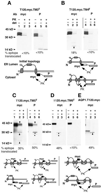
Figure 3.
Topological reorientation of TM3 in XO. Plasmids encoding the myc reporter at AQP1 residue T120 and the P reporter after TM3, TM4, TM5, or TM6 as indicated (A–D, respectively) were expressed in XO and digested with PK in the presence or absence of Triton X-100 (det) as described in MATERIALS AND METHODS. Samples were immunoprecipitated with anti-myc (myc) or anti-prolactin (P) antiserum and analyzed by SDS-PAGE. (E) Results for full-length AQP1.T120.myc. Upward arrowheads (B–E) indicate PK-protected polypeptide fragments generated by protease digestion. Downward arrowheads (C) indicate full-length chains in which cytosolic loops are not accessible to protease. Diagrams beneath each autoradiogram indicate potential topologies of myc and P epitopes deduced by PK digestion. Sites of PK digestion are indicated by arrows, whereas inaccessible sites are indicated by arrows blocked by lines. The topologies of myc and P reporters are derived from the accessibility of epitopes based on patterns of protected fragments.

