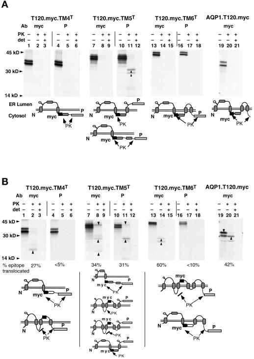
Figure 4.
In vitro AQP1 topology. Expression of T120.myc.TM4T, -TM5T, and -TM6T and AQP1.T120.myc in RRL and canine pancreas microsomes (A) or oocyte-derived ER membranes (B). Translation products were digested with PK and immunoprecipitated as in Figure 3. Upward arrowheads indicate polypeptide fragments protected from PK digestion in the absence, but not in the presence, of detergent. Potential topologies of epitopes indicated beneath autoradiograms are deduced from the protease accessibility of myc and P reporters. Sites of PK digestion are indicated.

