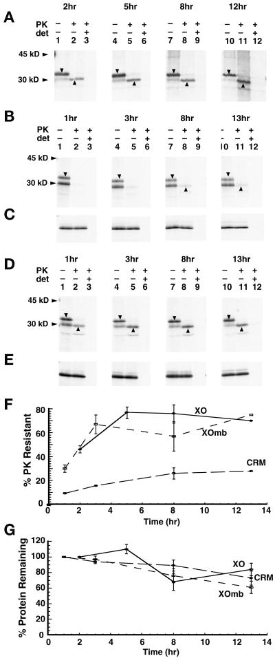
Figure 5.
Topological maturation of AQP1 in vivo and in vitro. Plasmids encoding AQP1.T120.myc were expressed in microinjected Xenopus oocytes (A), RRL supplemented with CRM (B), and RRL supplemented with ER-derived oocyte ER membranes (D) as described in MATERIALS AND METHODS. At the times indicated after RNA injection (A) or the initiation of in vitro translation (B–E), samples were digested with PK and immunoprecipitated as in Figures 3 and 4. Downward arrowheads indicate glycosylated full-length AQP1 chains. Upward arrowheads indicate the PK-protected AQP1 hydrophobic core. C and E show protease protection results of the secretory control protein, bovine prolactin, translated and incubated in CRM and XOmb, respectively. (F) Protease resistance of AQP1.T120.myc was determined by the fraction of glycosylated chains that had acquired PK resistance. (G) Total AQP protein at each time point. Results represent the average of three to four separate experiments ± SE.

