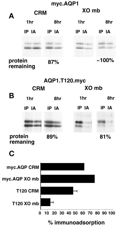
Figure 6.
Immunoadsorption of myc-tagged AQP1. Plasmids encoding the myc epitope at residue M1, myc.AQP1 (A), or residue T120, AQP1.T120.myc (B), were translated in RRL in the presence of CRM or XOmb for 1 h. After translation, immunoadsorption (IA) and immunoprecipitation (IP) were performed as described in MATERIALS AND METHODS. (C) Percent protein recovered by immunoadsorption relative to protein recovered by immunoprecipitation after the 8-h chase period. Total protein remaining at 8 h is indicated beneath the autoradiograms. Values indicate the average of two experiments for the control plasmid, myc.AQP1, and three experiments (±SE) for AQP1.T120.myc.

