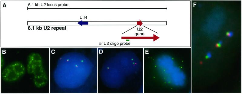
Figure 1.
Detection of RNA transcripts from the U2 locus. (A) Diagram of the 6.1-kb U2 repeat and positions of the U2 locus probe and 5′ U2 oligonucleotide probe. See Figure 5 for the sequence of the 5′ U2 oligonucleotide. (B) Distribution of U2 snRNA in HeLa nucleus detected with the U2-specific oligonucleotide. (C and D) HeLa cell nucleus hybridized for DNA (green) and RNA (red) with the 6.1-kb U2 gene locus probe shows gene foci without significant RNA signals associated or not associated with a DNA signal. There are three U2 loci in HeLa cells. (E) U2 locus RNA hybridization (green) in a metaphase cell. Numerous discrete foci are evident. (F) HeLa cell nucleus hybridized with the U2 locus probe for U2 DNA (green), RNA (red), and stained with an anti-coilin antibody to visualize CBs (blue). This nucleus shows a CB with each of the three gene locus foci as well as a separate RNA signal, possibly separated from the nearest gene locus.

