Figure 3.
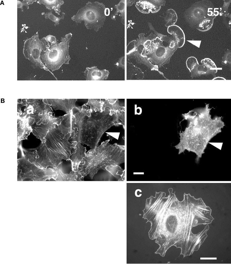
Lamellipodium formation after stimulation with PMA is Rac1 dependent. (A) GFP-actin–transfected B16 cells were plated on FN and treated with 100 ng/ml PMA at time 0′. Cells show intense ruffling and lamellipodium formation 55′ after addition of PMA (arrowheads). Bar, 40 μm. (B) B16 melanoma cells transiently transfected with N17Rac (dominant negative) were plated on FN and stimulated with 100 ng/ml PMA for 30 min. Fixed cells were stained for actin (a) and double labeled for N17Rac expression (b). In contrast to nontransfected cells, the N17Rac-transfected cell does not show stress fibers and lamellipodia (a, b). Bar, 20 μm. B16 cell cotransfected with GFP-actin and L61Rac (constitutively active) plated on FN (c) exhibits stress fibers and a smooth rim around the cell edge. Bar, 20 μm.

