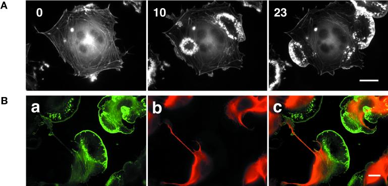
Figure 5.
PMA-induced lamellipodium formation in B16 cells pretreated with taxol. (A) Melanoma cells transfected with GFP-actin were plated on FN and treated with 10 μM taxol for 1 h prior to 100 ng/ml PMA at time 0′. Subsequent images show circular actin ruffles and lamellipodium formation after addition of PMA (time 10′ and 23′). Bar, 20 μm. (B) Tubulin and actin distribution in cells treated with taxol and PMA. GFP-actin–expressing cells were plated on FN and treated subsequently with taxol and PMA. Cells were then fixed and stained for tubulin. a, actin distribution; b, tubulin distribution; and c, overlay of tubulin (red) and actin (green) within the cell. Note the cells show lamella and actin-rich lamellipodium (a, c) without any tubular structures (b, c). Bar, 20 μm.

