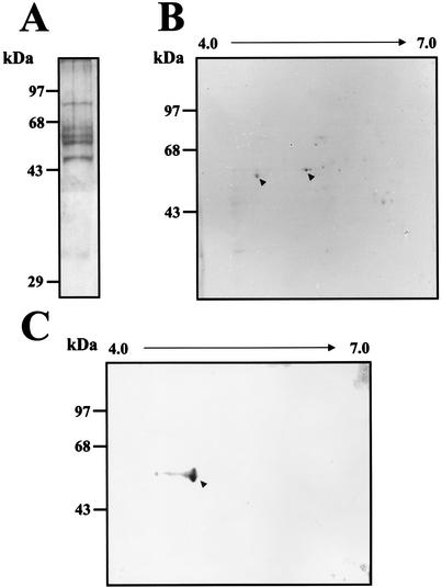
FIG. 8.
Proteins present in the CI complex. (A) After a mobility shift assay, the CI from infected S10 extract was cut from the native gel, electroeluted, and then subjected to SDS-10% PAGE. The proteins were stained using Coomassie blue. (B) The proteins obtained from the CI were separated through two-dimensional electrophoresis. The proteins indicated with arrowheads were cut and sequenced. (C) Western blot assay of the proteins present in the CI complex using an anticalreticulin polyclonal antibody. Molecular mass markers are indicated on the left. The arrowhead indicates the calreticulin protein.

