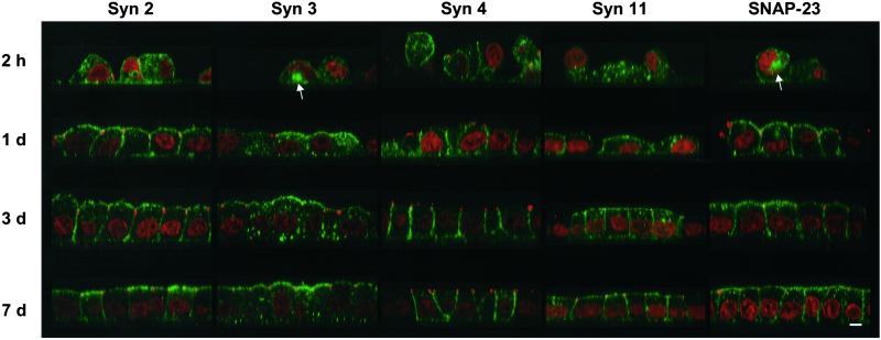
Figure 3.
All plasma membrane t-SNAREs relocalize from intracellular compartments to their final plasma membrane domains during development of MDCK cells into a polarized monolayer. MDCK cells stably expressing syntaxins 2, 3, 4, and 11 and SNAP-23 were seeded at high density onto polycarbonate filters. At the indicated times, the cells were fixed and stained with affinity-purified antibodies against the respective SNAREs (green) as well as an antibody against the tight junction protein ZO-1 (red). Nuclei were stained with propidium iodide (red). Confocal optical sections through the monolayers are shown with the apical side on top. Once the cells have established contacts, the tight junctions can be seen as red dots at the junction between the apical and basolateral plasma membranes. At the earliest time, large intracellular vacuoles are occasionally detected (arrows), most frequently with syntaxin 3 and SNAP-23. While the distribution of all studied SNAREs is predominantly intracellular at 2 h, it shifts to a predominantly plasma membrane localization during the course of 7 d. Note that starting at d 1, syntaxin 3 is always excluded from the basolateral plasma membrane, whereas syntaxin 4 is always excluded from the apical plasma membrane. Bar, 5 μm.

