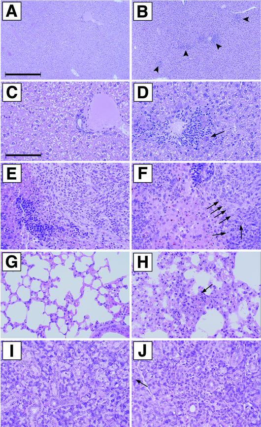
FIG.5.
Histological examination of livers, spleens, lungs, and salivary glands from mice infected with enhancerless MCMV. Shown are sections from livers (A to D), spleens (E and F), lungs (G and H), and salivary glands (I and J) of CB17 SCID mice 14 days after infection with 6 × 10⁵ PFU of tissue culture-passaged MCMVdE::Luc (A, C, E, G, and I) or MCMVdE::Luc-rev (B, D, F, H, and J). (A, C, E, G, and I) No lesions present. (B) Multifocal inflammatory foci (marked by arrowheads). (D) Note cytomegalic cell with inclusion (arrow). The number of inflammatory foci present in liver sections from MCMVdE::Luc-rev-infected animals was 23 ± 10 per mm2. (A and B) Overview; (C and D) details of portions of A and B, respectively. (F, H, and J) Note the cytomegalic cells (indicated by arrows). (F) Neutrophilic infiltrate in splenic white pulp. (H) Lung alveolar septal thickening with fibrin and inflammatory cells. (J) Salivary gland focal necrosis with inflammation. Bars, 400 (for A and B) and 100 (for C through J) μm. Magnification, ×25 (A and B) and ×100 (C through J).

