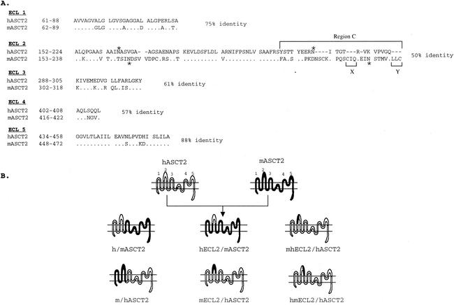
FIG. 1.
Construction of hASCT2/mASCT2 chimeras. (A) Amino acid sequence comparison of the putative ECL regions of hASCT2 and mASCT2. Numbers at the left of the sequences correspond to the positions of the first and last amino acids shown. Dots indicate amino acid identity. Deletions in sequences are indicated by dashes. N-glycosylation sites are indicated by asterisks. Numbers at the right of the sequences indicate the percent amino acid identity. (B) The topologies and the nomenclatures of the wild-type and chimeric ASCT2 proteins. The representation on the top shows the putative topology of the hASCT2 and mASCT2 cell surface receptors; the putative ECLs are numbered 1 through 5.

