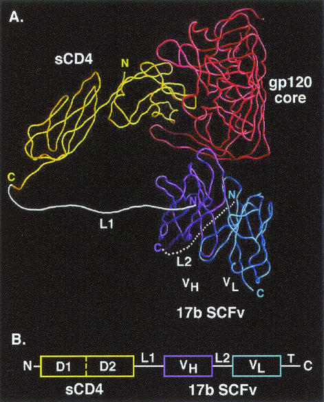
FIG. 1.
sCD4-17b design. (A) Predicted structure of sCD4-17b interacting with gp120. A Cα worm diagram showing the trimeric complex containing the gp120 core (red), two-domain sCD4 (yellow), and the 17b SCFv (VH, purple; VL, blue) was derived from the coordinates of the published X-ray crystallographic complex (18), using the GRASP program (27). The L2 linker is depicted at the back of the complex. The thrombin cleavage site followed by the 6-His tag is not shown. (B) Schematic of the sCD4-17b genetic construct (not drawn to scale), including the CD4 leader at the N terminus and the thrombin cleavage site followed by 6 histidne residues (T) at the C terminus.

