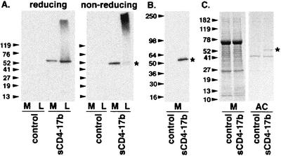
FIG. 2.
Expression and affinity chromatography of sCD4-17b. BS-C-1 cells were infected with WR (control) or vCD-3 (sCD4-17b). SDS-PAGE was used to resolve samples of media (M) and corresponding detergent lysates (L). In each experiment, arrowheads indicate the migration according to molecular size standards (in kilodaltons), and an asterisk indicates the position of the sCD4-17b band. (A) Immunoblot analysis with anti-CD4 polyclonal antisera, on reducing (left panel) and nonreducing (right panel) gels. (B) Immunoblot analysis of concentrated media with a MAb against the His tag. (C) Coomassie blue staining of concentrated media (M) and affinity chromatography-enriched (AC) fractions. Each lane in panels A and B represents material from equivalent numbers of cells; to facilitate visualization in panel C, the affinity chromatography-enriched samples represent material from three times the number of cells as the concentrated media samples.

