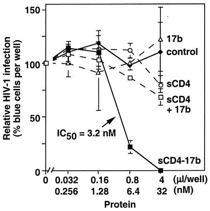
FIG. 3.
Neutralizing activity of sCD4-17b against a primary HIV-1 isolate. The Ba-L strain was preincubated with increasing volumes of concentrated media from BS-C-1 cells infected with either vCD-3 (sCD4-17b, corresponding to the indicated concentrations) or WR (none); equivalent virus samples were preincubated with increasing amounts of the purified protein sCD4, 17b IgG, or sCD4 plus 17b IgG. The infectivity assay was performed with MAGI-CCR5 cells. Results are expressed as the percentage of blue cells compared to the level (taken as 100%) in samples incubated with no additions (which gave 108 blue cells per well). Error bars indicate standard deviations of duplicate samples. On the x axis, the “μl/well” label refers to the volumes of concentrated media from vaccinia virus-infected cells; the “nM” label indicates the corresponding concentrations of sCD4-17b in the concentrated medium of vCD-3-infected cells, as well as the concentration of each purified protein.

