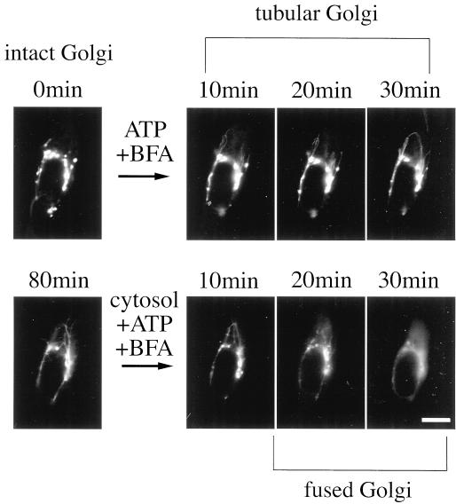
Figure 3.
Morphological dissection of the BFA-induced Golgi disassembly process in a single semi-intact cell. Semi-intact cells were incubated with BFA/ATP at 30°C for 80 min to generate the Golgi tubules. After being washed, the cells were further incubated with BFA/cytosol/ATP for 80 min. The sample was imaged at 5-min intervals with the use of a conventional fluorescence microscope. The Golgi tubules fused with the ER networks only in the presence of both exogenous cytosol and ATP. Bar, 10 μm.

